Getting pregnant
Pregnancy beginning
Pregnancy monitoring
Childbirth
Special cases
Post-partum immédiat
Post natal consultation
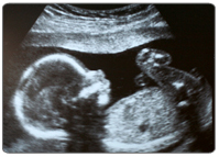
Fetal ultrasound
You will be offered 3 ultrasounds, 1 per trimester.
Ultrasound allows to get information no other examination can give: exact date of pregnancy beginning, fetus number, fetus growth, blood flow between the mother and its fetus (Doppler ultrasound) and fetus morphology.
These are not mandatory examinations. Sonography can reveal birth defect. Today’s technology is quite advanced, but ultrasound is not perfect and abnormalities may not being detected.
On another hand, some characteristics observed during ultrasound may suggest birth defect while there is none. In this case, ultrasound can be anxiety-provoking, either for you or your spouse. In case of doubt, supplementary exam will be realized (as amniocentesis, blood test or MRI) and control exam will be suggested to you.
In practice, ultrasound is realized either by the doctor or the midwife. It is completely painless and without any either for the mother or her baby.
A small handheld device (transducer) is placed onto your skin and moved over your womb. A lubricating gel is put onto your skin to allow transducer to move slowly. Transducer is connected to a computer and a monitor, displaying still pictures and movements of the fetus.
In some situations, a thin transducer introduced into the vagina will be used, in order to visualize some less accessible parts of the fetus or its “annexes” (placenta, membranes, amniotic fluid).
There is no need to have empty stomach. For the first ultrasound – and sometimes for the followings – you may need to have a full bladder during the exam.
Important things to do:
- Do not apply any moisturizing cream 48 hours before the ultrasound
- Avoid any other children and too many persons around you and your spouse during the exam.
Last update: 10/2/2013